Diabetes mellitus affects 194 million people worldwide and is expected to increase in prevalence to 439 million by the year 2030. Foot ulceration is a major complications which occurs in as many as 15-25% of type 1 and 2 patients with diabetes mellitus over their lifetime. If left untreated, foot ulcers may become infected and develop deep tissue necrosis which may require amputation. It has also been estimated that foot ulcers precede roughly 85% of all lower extremity amputations in patients with diabetes mellitus and more than 88,000 lower limb amputations are performed annually on diabetic patients in the United States. The cost of foot disorder diagnosis and management is estimated to be over $6 billion dollars annually in the United States. Furthermore, it has been suggested that 40% to 85% of diabetic foot amputations can be avoided with early detection and preventive techniques such as offloading and improved hygiene. Automated early identification of tissue at risk of ulcerating may enable directed care thereby reducing the incidence of foot ulceration and amputation. This study proposes to use hyperspectral imaging as a screening tool for detecting forming diabetic foot ulcers before tissue damage becomes apparent to a care-giver in a clinical setting.
Tissue oximetry measurements were performed during several visits with hyperspectral imaging of the feet in type 1 and 2 diabetes mellitus subjects that were at risk for foot ulceration.
- Enrolled 153 patients with Type 1 and 2 Diabetes Mellitus at risk of developing foot ulcers.
- Institutional Review Board: Olive View-UCLA IRB #05H-609300
- Followed patients over a 24-week period for 11 total visits.
- After signing the informed consent form, hyperspectral imaging of the feet was performed at each visit and preulcer sites analyzed retrospectively.
- Patients were followed for 18 months.
- The clinical study was performed by Dr. Aksone Nouvong, DPM
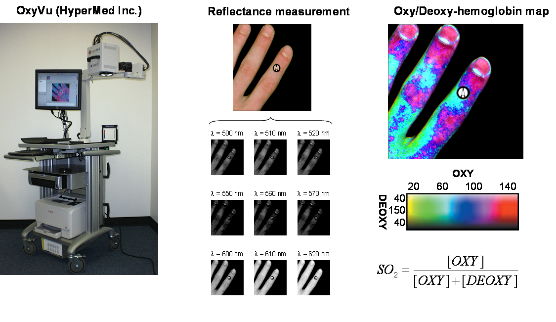
Oximetry Based Algorithm and Hypothesis
The data was retrospectively analyzed at 21 sites that ulcerate during the course of the study and an ulceration prediction index was developed. We calculated the maximum absolute difference (
.png)
Scatter plot showing values of MAD(OXY) and MAD(DEOXY) determined for 21 affected and 21 contralateral sites from diabetic subjects who developed foot ulcers and from 100 random sites from diabetic subjects in the comparative groups. Values from affected areas were found to fall inside the grey regions where |MAD(OXY)|> 18 and |MAD(DEOXY)|> 5.8 with a p-value of less than 0.0001.
.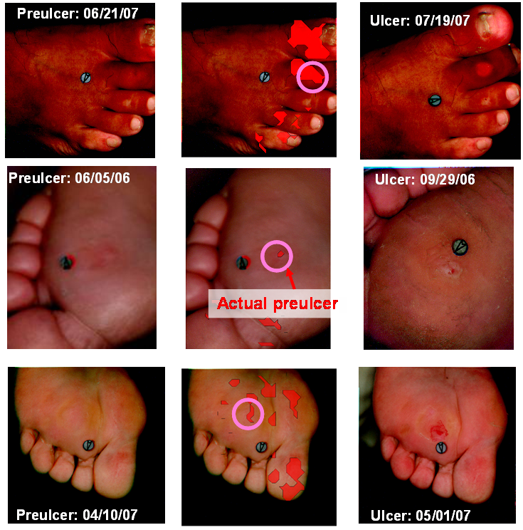
Composite image of three diabetic feet where the red overlay indicates that the maximum absolute differences in oxyhemoglobin and deoxyhemoglobin are such that |MAD(OXY)|> 18 and |MAD(DEOXY)|> 5.8. The approximate location of subsequent ulceration is circled.
D. Yudovsky, A. Nouvong, K. Schomacker, and L. Pilon, 2011. Monitoring Temporal Development and Healing of Diabetic Foot Ulcer Using Hyperspectral Imaging, Journal of Biophotonics, Vol. 4, No. 7-8, pp. 565-576. doi:10.1002/jbio.201000117 pdf
D. Yudovsky and L. Pilon, 2011. Retrieving Skin Properties From In Vivo Diffuse Reflectance Measurements on Human Skin, Journal of Biophotonics, Vol. 4, No.5, pp.305-314, doi:10.1002/jbio.201000069 pdf
D. Yudovsky, A. Nouvong, K. Schomacker, and L. Pilon, 2011. Assessing Diabetic Foot Ulcer Development Risk with Hyperspectral Tissue Oximetry, Journal of Biomedical Optics, Vol.16, No.2, 026009. doi: doi:10.1117/1.3535592 pdf
D. Yudovsky and L. Pilon, 2010. Modeling of Local Excitation Fluence Rate and Florescence Emission in Absorbing and Strongly Scattering Multilayered Media. Applied Optics, Vol. 49, No. 31, pp. 6072-6084. doi:10.1364/AO.49.006072 pdf
D. Yudovsky , A. Nouvong, and L. Pilon, 2010. Hyperspectral Imaging for Diabetic Foot Wound Care, Journal of Diabetes Science and Technology, Vol.4, No.5, pp. 1099-1113. pdf