This study combines non-invasive hyperspectral imaging with our experimentally validated skin optical model (Yudovsky and Pilon, 2009) and inverse algorithm (Yudovsky and Pilon, 2010) to monitor diabetic feet of two representative patients. It aims to observe temporal changes in local epidermal thickness and oxyhemoglobin concentration and to gain insight into the progression of foot ulcer formation and healing. Foot ulceration is a debilitating comorbidity of diabetes that may result in loss of mobility and amputation. Inflammation and necrosis preempt ulceration and can result in changes in the skin prior to ulceration and during ulcer healing that affect oxygen delivery and consumption.
Hyperspectral imaging of the feet of two diabetic patients was performed before, during, and after they developed foot ulcer. Previous studies of diabetic foot ulcer formation using hyperspectral imaging limited their analysis to circulatory changes such as difference in oxyhemoglobin and deoxyhemoglobin concentrations obtained from hyperspectral images collected immediately prior to foot ulcer formation or immediately following ulcer healing. The present study examined the temporal changes observed before the ulcer became apparent to the naked eye until it healed and closed. Variables of interest were local epidermal thickness, dehoxyhemoglobin, oxyhemoglobin, as well as oxygen saturation.
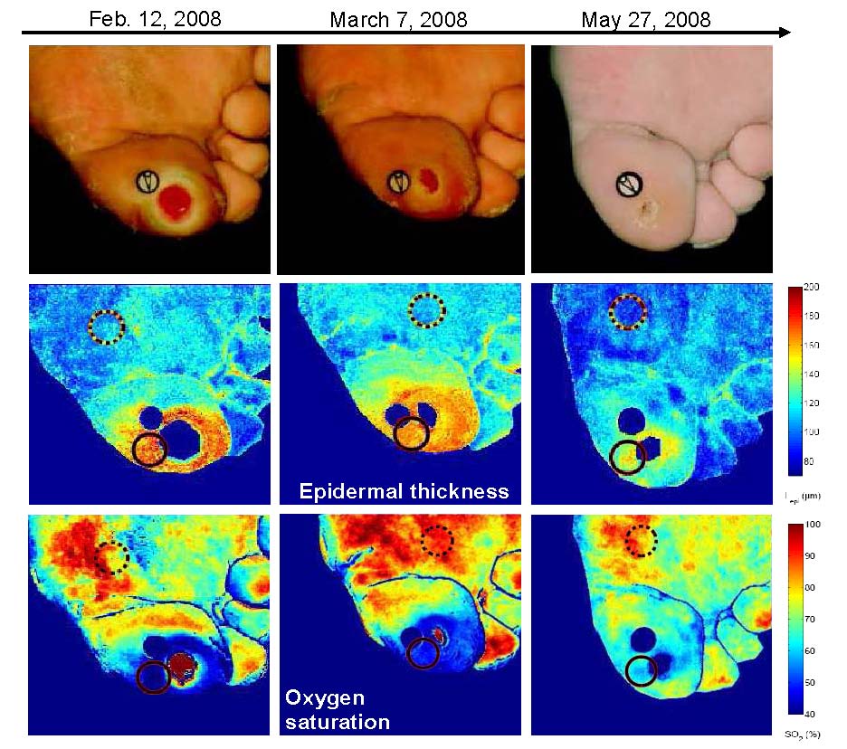
This study showed that epidermal thickening and decrease in oxyhemoglobin concentration can be detected non-invasively prior to ulceration at pre-ulcerative sites. The algorithm was also able to observe reduction in the epidermal thickness combined with an increase in oxyhemoglobin concentration around the ulcer as it healed and closed. This methodology can be used for early prediction of diabetic foot ulceration in a clinical setting.
D. Yudovsky, A. Nouvong, K. Schomacker, and L. Pilon, 2011. Monitoring Temporal Development and Healing of Diabetic Foot Ulcer Using Hyperspectral Imaging, Journal of Biophotonics, Vol. 4, No. 7–8, pp. 565–576. doi: 10.1002/jbio.201000117.
D. Yudovsky and L. Pilon, 2011. Retrieving Skin Properties From In Vivo Diffuse Reflectance Measurements on Human Skin, Journal of Biophotonics, Vol. 4, No.5, pp.305-314, doi: 10.1002/jbio.201000069.
D. Yudovsky, A. Nouvong, K. Schomacker, and L. Pilon, 2011. Assessing Diabetic Foot Ulcer Development Risk with Hyperspectral Tissue Oximetry, Journal of Biomedical Optics, Vol.16, No.2, 026009. doi: 10.1117/1.3535592.
D. Yudovsky and L. Pilon, 2010. Modeling of Local Excitation Fluence Rate and Florescence Emission in Absorbing and Strongly Scattering Multilayered Media. Applied Optics, Vol. 49, No. 31, pp. 6072–6084. doi: 10.1364/AO.49.006072
D. Yudovsky , A. Nouvong, and L. Pilon, 2010. Hyperspectral Imaging for Diabetic Foot Wound Care, Journal of Diabetes Science and Technology, Vol.4, No.5, pp. 1099-1113.
D. Yudovsky and L. Pilon, 2009. Simple and Accurate Expressions for Diffuse Reflectance of Semi-Infinite and Two-Layer Absorbing and Scattering Media, Applied Optics, Vol. 48, no. 35, pp. 6670-6683. Selected to appear in The Virtual Journal for Biomedical Optics,Vol. 5, No.1, January 4, 2010. doi: 10.1364/AO.48.006670